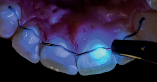
Low invasive fluorescence guided dentistry
Techniques and methods that allow the preservation of dental tissues.
Low-invasive dentistry encompasses a philosophy that integrates prevention, remineralization, and minimal intervention for the placement and replacement of dental restorations. Any treatment based on the least invasive restorative approach is to remove the minimum amount of healthy tissue. Thus, the use of techniques and methods that allow the preservation of dental tissues is the desire to realize this work. Furthermore, the diverse characteristics and optical properties such as translucency, opalescence, and fluorescence of the dental hard tissues that we face in the removal and preparation of restorations, give us the necessary information to not go beyond our objective, thus trying to preserve and conserve their natural and aesthetic structure.
The fluorescence of natural teeth is of great interest to the clinician because it is associated, among other things, with a dental vitality effect, the absence of which is evident in greyish restorations. In today's world, surrounded by the artificial light influence of UV radiation (blacklight in bars, fluorescent tubes in studios, game centers, discotheques), as well as frequent exposure to the sun effect, all these factors influence the level of perceived vitality of restorations and, therefore, their naturalness. Human teeth are naturally fluorescent or auto-fluorescent, as UV rays are easily absorbed by the luminescent components in dental tissues.
So, how strong would fluorescence and UV be in minimally invasive restorative dentistry? What if we want to copy the characteristics of our dental issues? We must consider the presence of organic substances, both dentin and enamel. These structures emit different degrees of fluorescence, and distinguishing them will preserve the most significant amount of tissue and use resins whose property of casting such a light characteristic is similar to the tooth to be restored.
How does fluorescence occur in dental tissues?
When UV light A of the non-visible spectrum (between 350 and 400 nm) strikes dental tissues, it is initially absorbed and then emitted in the visible range at longer wavelengths (between 410 and 500 nm) (5). The teeth generally appear intensely white and bluish under UV light, making them appear bright and vital. Dentin and enamel are dental structures with fluorescent properties. However, dentin is more accentuated due to more organic components, such as proteins and photosensitive collagen fibers. In enamel, the low content of organic ingredients is responsible for its lower fluorescence. This is also the reason why the cervical area of the tooth fluoresces more than the incisal area.
The fluorescence of aesthetic restorative materials
The use of new aesthetic materials, such as composite resins and ceramics that meet the optical characteristics similar to the tooth structure, has led manufacturers to incorporate various chemicals and minerals such as fluorophores type europium oxide, cerium, terbium, and samarium or so-called rare-earth to achieve the naturalness of dental restorations. However, advances in the composition of new dental resins are a challenge for manufacturers to reproduce the optical properties of dental tissues satisfactorily due to factors such as ambient light conditions and the dental aging process, among others. In the case of dental ceramics, there are many differences in composition, and it also depends on their sintering processes and cycles and the effects of temperature, which reduce and affect the intensity of fluorescence. Traditional zirconium is a non-fluorescent material. Different products have been commercialized to improve the optical conventional zirconium behavior oxides, such as coatings (to be applied on their surface) and fluorescence modifying liquids to immerse the zirconium oxide parts before sintering. Despite the limited research on the behaviofluorescence behavioresthetic materials, manufacturing companies are increasingly interested in developing this field.
Material and methods for the use of fluorescence-assisted identification
The assisted or fluorescence-induced identification technique in dental practice consists of using an auxiliary UV A light whose spectrum is between 360-410 nm. Currently, the search to design an electronic device of a different and complementary diagnostic nature when evaluating the aesthetics of restorations has been the initial motivation to create K-Lite (Smileline-Switzerland & Dr. Katherine Losada), a lightweight double LED (White Light for use in translumination and violet light to induce fluorescence) electronic device with a 3mm diameter fiberglass guide and without wires, for daily use whose wavelength close to 400 nm has proven to be effective in easy identification of enamel and dentin dental tissues (increased fluorescence, brightness, and white-blue intensity), essential when making less invasive enamel preparations.
- Identification of enamel caries and initial white lesions due to decreased fluorescence from demineralization. Also, the differentiation of affected and infected dentin in the presence of certain bacteria, whose by-product is porphyrins, emits intense reddish-pink fluorescence in the UV light type A.
- Simple recognition of calculus or tartar, both supra and subgingival (emits red-orange fluorescence).
- Distinguishing and identifying aesthetic restorative materials that are in contact with natural dental underlying tissues (to assist in the removal of old and defective restorations and/or dental adhesives such as orthodontic anchors)
- Control the margins of pre-existing restorations without the need for routine and decrease the use of radiographs for location and extension.
Summary
In daily dental practice, we must consider that we commonly evaluate restorations and dental tissues in an artificial environment that simulates natural light. However, this lighting does not allow us to identify the enamel and dentin for minimal tissue preparation. The margins of tooth-colored restorations, the limit of dental adhesives, and their possible excesses are not simple to perceive. The main reasons for the patient to attend a dental consultation are the replacement of repairs, the use of aesthetic materials, the placement of transparent aligners, the identification of the initial carious lesions, and the removal of dental calculus. Using fluorescence through an ultraviolet light device would benefit the removal of such materials by avoiding excessive removal, iatrogeny, and/or involuntary contact with the underlying healthy dental tissues.
Currently, tests performed on clinical cases and the compliance and certification of K-Lite as a device for dental and medical use have shown effectiveness in its daily use as an easy and routine tool to assist the clinician during dental diagnosis and treatment.

